Histopathology
- Research associate:
- Postdoctoral fellow:
- Laboratory technicians:
The Unit delivers end-to-end histopathological workflows, from necropsy and tissue collection to advanced staining and expert evaluation. Standard processing includes fixation, paraffin embedding, sectioning, and H&E staining, complemented by a broad panel of special stains and immunohistochemistry for detailed tissue characterization. A key strength of the platform is interpretation by trained veterinary and clinical pathologists, ensuring consistent and biologically meaningful evaluation of tissue alterations. Structured reports with high-quality image documentation support robust phenotype definition and cross-study comparability. These workflows enable integration of tissue-level findings with molecular, physiological, and behavioral data, supporting both mechanistic understanding and translational preclinical applications.
Standard services Gross morphology & necropsy
Complete necropsy of mouse and rat models performed by trained veterinary pathologists, including standardized documentation of macroscopic findings and systematic organ collection.
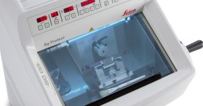
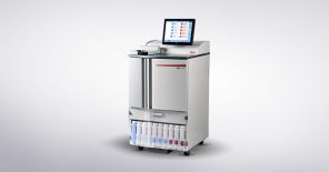
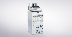
Standard services Tissue processing & embedding
Automated fixation, paraffin embedding, and processing workflows ensuring high reproducibility and consistency across studies, including optional decalcification and frozen tissue preparation.
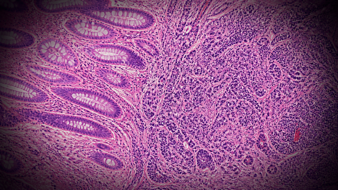
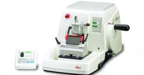
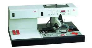
Standard services Sectioning & routine staining (H&E)
High-throughput sectioning and automated hematoxylin and eosin staining as the baseline for morphological assessment across all projects.
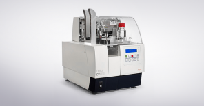
Standard Services Special stains panel
Special stains are used to differentiate key biological constituents, including lipids, carbohydrates, amyloid deposits, and connective tissue, and therefore represent a powerful tool for detection, scoring, and monitoring of diverse pathological processes such as fibrosis, steatosis, and amyloidosis.
In experimental and preclinical research, special stains provide a robust and often underutilized complement to immunohistochemistry. In many contexts, they offer a cost-effective and highly reproducible alternative for assessing tissue architecture and composition, particularly in large-scale or screening-oriented studies.
The Unit maintains a broad portfolio of validated histochemical stains and supports implementation of additional protocols upon request.
Available stains include:
Alcian Blue, Alcian Blue + PAS, Congo Red, Giemsa, Gram staining, Masson’s Trichrome, NASDCL, New Methylene Blue, Nissl, Oil Red O, PAS, Picrosirius Red, Prussian Blue, Reticulin, TRAP.
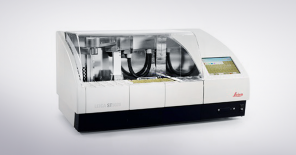
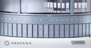
Standard Services Immunohistochemistry (IHC) – validated workflows
Standardized immunohistochemistry pipelines using automated platforms, with validated protocols ensuring reproducibility across experiments. The list of validated antibodies [link].
Do you have questions? Ask us
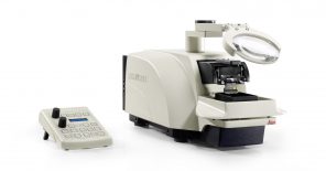
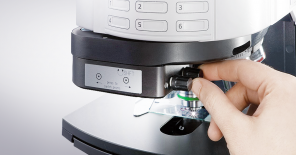
Standard services Slide scanning & digital pathology
High-resolution brightfield and fluorescence slide scanning enabling digital archiving, remote evaluation, and quantitative image analysis.
Do you have questions? Ask us
Project Specific Assays Quantitative histopathology & scoring frameworks
Custom scoring systems for lesion grading, fibrosis staging, inflammation quantification, or tumor characterization, tailored to specific disease models and aligned with preclinical or regulatory standards.
Project Specific Assays Advanced immunophenotyping & spatial biology
Design and optimization of multiplex or targeted IHC workflows to characterize cellular composition, immune infiltration, and pathway activation in tissues.
Project Specific Assays Tissue-based efficacy readouts in preclinical studies
Histopathological evaluation of treatment effects, including toxicity assessment, target organ response, and disease modification across longitudinal or endpoint studies.
Project Specific Assays Integrated morphology–omics workflows
Preparation of tissue samples compatible with downstream molecular analyses (RNA, proteomics, spatial profiling), enabling direct correlation between morphology and molecular signatures.
Project Specific Assays Custom sample processing pipelines (non-standard materials)
Tailored workflows for organoids, cell cultures, biopolymers, rare tissues, or non-rodent samples, including protocol optimization for fixation, embedding, and staining.
Project Specific Assays Reproductive & endocrine histopathology
Histological evaluation of reproductive organs, including estrous cycle staging (e.g. Shorr staining) and integration with hormonal profiling.
Do you have questions? Ask us
Technology platforms
Histopathology unit was upgraded with the support from OP RDE project CZ.02.1.01/0.0/0.0/18_046/0015861 CCP Infrastructure Upgrade II.
