Vision
- Laboratory technician:
- PhD student:
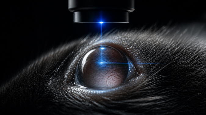
High-resolution SD-OCT enables quantitative, layer-resolved analysis of retinal morphology, including thickness, structural integrity, and optic disc architecture. OCT angiography (OCT-A) further provides detailed assessment of retinal and choroidal vascular networks, including vessel density, branching, and perfusion characteristics. These approaches allow sensitive detection of disease-associated changes and their progression over time. Functional relevance is assessed by electroretinography (ERG), measuring light-evoked responses of retinal cell populations. Together, structural and functional readouts support longitudinal studies and evaluation of therapeutic interventions, including preclinical models of retinal degeneration, vascular pathology, and neurodegenerative processes.
Comprehensive Vision Phenotyping Pipeline

Standard services Retinal Imaging and Vascular Analysis (SD-OCT & OCT-A)
High-resolution spectral-domain OCT enables non-invasive, layer-resolved imaging of retinal structure, including quantitative assessment of retinal thickness, integrity, and optic disc morphology. OCT angiography (OCT-A) provides detailed visualization of retinal and choroidal vascular networks, allowing analysis of vessel density, architecture, and microvascular changes. These approaches enable sensitive detection of retinal degeneration, edema, and vascular pathology, and are particularly suited for longitudinal studies and monitoring of disease progression or therapeutic response.
Standard services Advanced Retinal and Vascular Imaging (FA & ICG)
Fluorescein angiography (FA) and indocyanine green angiography (ICG) provide complementary assessment of retinal and choroidal circulation. These methods enable detection of vascular leakage, neovascularization, and perfusion abnormalities that cannot be fully captured by non-invasive imaging alone. Due to their invasive nature, these techniques are primarily used as targeted follow-up tools in disease models requiring detailed vascular characterization.
Standard services Anterior Segment Imaging (Pentacam HR)
High-resolution anterior segment tomography enables quantitative analysis of corneal structure, curvature, and anterior chamber geometry. This modality is particularly relevant for experimental models of corneal pathology, structural abnormalities, and intervention studies, providing reproducible morphological readouts adapted for preclinical research.
Standard services Intraocular Pressure (IOP) Measurement
Non-invasive measurement of intraocular pressure using rebound tonometry provides a sensitive readout of ocular physiology and is particularly relevant in glaucoma models and studies of ocular hypertension. The method allows repeated measurements under minimal stress conditions, supporting longitudinal study designs.
Standard services Retinal Function Assessment (Electroretinography, ERG)
Electroretinography (ERG) enables functional evaluation of retinal cell populations by measuring light-evoked electrical responses. Standard protocols allow assessment of rod and cone function, as well as inner retinal signaling under scotopic and photopic conditions. ERG provides critical functional validation of structural findings and enables interpretation of the physiological impact of morphological abnormalities in disease models and therapeutic studies.
Do you have questions? Ask us
project specific assays Longitudinal Retinal Phenotyping Pipeline
The Unit provides integrated longitudinal assessment of retinal structure and function by combining SD-OCT, OCT angiography, and electroretinography (ERG). This approach enables sensitive detection and tracking of disease progression, including neurodegeneration, vascular remodeling, and retinal dysfunction. Repeated non-invasive measurements allow high-resolution temporal analysis of phenotypic changes and therapeutic response, making the platform particularly suitable for preclinical intervention studies.
project specific assays Functional ERG Challenge Paradigms
Advanced electroretinography protocols enable detailed functional characterization of retinal cell populations under defined stimulation conditions. These include dark- and light-adapted responses, flicker stimulation, and customized protocols assessing photoreceptor and inner retinal function. Challenge-based paradigms allow detection of subtle functional impairments and provide mechanistic insight into disease processes and treatment effects beyond standard structural imaging.
project specific assays Ocular Gene Therapy and Targeted Intervention Readouts
The Unit supports evaluation of ocular gene therapy and targeted intervention strategies through integrated structural and functional readouts. Retinal imaging (SD-OCT, OCT-A) combined with ERG enables comprehensive assessment of treatment efficacy, including tissue integrity, vascular response, and functional recovery. These workflows can be directly linked to image-guided intraocular administration of therapeutic agents, viral vectors, or cells in preclinical studies. (see Cardiovascular Unit), providing a complete preclinical pipeline from targeted intervention to quantitative phenotypic outcome.
Do you have questions? Ask us

Vision unit was upgraded with the support from OP RDE project CZ.02.1.01/0.0/0.0/18_046/0015861 CCP Infrastructure Upgrade II
