Cardiovascular
- Research associate:
- Research assistant:
- Laboratory technicians:
Beyond routine cardiovascular assessment, the Unit integrates imaging and physiological monitoring into broader preclinical phenotyping pipelines. High-resolution ultrasound, Doppler, and photoacoustic imaging are combined with ECG, telemetry, and blood pressure measurements to provide comprehensive characterization of cardiac and systemic physiology. The platform supports studies from embryonic development to adult disease models, including longitudinal and stress-based paradigms. This integrated approach enables detailed evaluation of disease mechanisms, functional reserve, and therapeutic responses across cardiovascular and multi-organ contexts.

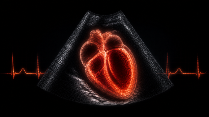
Standard services Echocardiography and Cardiovascular Imaging
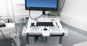
The Unit provides high-resolution in vivo echocardiography for detailed assessment of cardiac structure and function, including chamber dimensions, wall thickness, and valvular function. Functional analysis includes systolic and diastolic parameters, ejection fraction, fractional shortening, and myocardial strain as an early biomarker of dysfunction. Advanced ultrasound modalities, including Doppler, 3D/4D imaging, and ECG-gated acquisition, enable comprehensive evaluation of cardiac mechanics, vascular flow, and structural abnormalities in both adult and developmental models.
Standard services Electrocardiography (ECG) and Cardiac Electrical Function
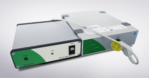
Non-invasive ECG monitoring enables detailed analysis of cardiac electrical activity under anesthetized or conscious, or freely moving conditions. Measurements include heart rate, conduction intervals, and arrhythmia detection, supporting studies of electrophysiology, disease models, and pharmacological interventions. Long-term telemetry allows continuous monitoring under physiological conditions, providing high-fidelity data for longitudinal and behavioral studies.
Standard services Blood Pressure and Hemodynamic Assessment
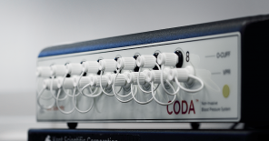
Non-invasive blood pressure measurement using high-throughput tail-cuff systems enables accurate assessment of systolic and diastolic pressure in conscious animals. These measurements support cardiovascular phenotyping in genetic, pharmacological, and environmental intervention studies.
Standard services Functional Cardiac Stress Testing
Cardiac function and physiological reserve can be assessed using controlled stress paradigms, including treadmill-based exercise testing and pharmacological stimulation (e.g. dobutamine challenge). These approaches enable evaluation of cardiac performance under increased workload and provide sensitive detection of subclinical phenotypes.
Standard services Developmental and Fetal Cardiovascular Imaging
The Unit supports ultrasound-based assessment of cardiovascular development, including in utero imaging of embryos and analysis of cardiac structure and blood flow dynamics. These workflows enable investigation of developmental phenotypes and early functional abnormalities.
Do you have questions? Ask us
Project Specific Assays Advanced Cardiovascular Imaging and Functional Analysis
Customized imaging workflows include photoacoustic analysis of tissue oxygenation, myocardial perfusion, and ischemia, as well as advanced strain-based assessment of cardiac mechanics. These approaches provide deeper insight into disease progression and therapeutic response.
Project Specific Assays Oncology and Systemic Imaging Applications
Ultrasound and photoacoustic imaging enable detailed analysis of tumor growth, vascularization, and perfusion, including longitudinal monitoring of therapy response. These workflows support both cardiovascular and oncology-oriented research.
Project Specific Assays Image-Guided Interventions and Targeted Delivery
Real-time ultrasound-guided injection enables precise delivery of cells, viral vectors, or therapeutic agents into defined anatomical locations, supporting advanced experimental designs including gene therapy and cell-based interventions.
Project Specific Assays Ex Vivo Cardiac Models and Cellular Assays:
The Unit provides isolation of primary cardiomyocytes using Langendorff perfusion and supports functional assays including contractility, electrophysiology, and calcium dynamics. These models enable mechanistic studies complementing in vivo phenotyping.
Project Specific Assays Multiorgan and Cross-System Sonography
Customized imaging protocols can be applied to multiple organs, including brain, liver, kidney, and reproductive tissues, enabling integrated assessment of systemic physiology and disease processes beyond the cardiovascular system.
Project Specific Assays Method Development and Advanced Imaging Engineering
The platform supports development of custom ultrasound acquisition protocols and advanced imaging approaches, enabling exploration of novel methodologies and adaptation to specific experimental needs.
Do you have questions? Ask us
Cardiovascular unit was upgraded and expanded with the support from OP RDE project CZ.02.1.01/0.0/0.0/18_046/0015861 CCP Infrastructure Upgrade II in the years 2020 – 2022 and currently it is being upgraded from the OP JAC project CZ.02.01.01/00/23_015/0008189 Upgrade of the large research infrastructure CCP III.